Prokaryotic-Bacteria-Cell-Parts| Notes by UK Sir | Cell Bio- 5
Prokaryotic-Bacteria-Cell-Parts
a) Bacterial cell Parts:
Want to know about Shape size of Bacterial cell, Gram staining Click the link below :
Want to know about Ultra Structure of Bacterial cell Click the link below :
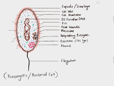
Mesosome:
-
it
was Discovered by F. James 1960
-
It
is Ingrowth of plasma membrane, finger
like projection
-
In
mesosome Vesicles, tubules, lamellae present
-
It
is of two types- Septal and Lateral
 |
| Septal mesosome |
-
i)
Septal: Connected with nucleoid and membrane,
help in replication of Nucleoid and
cell division.
-
ii) Lateral : don’t connected with nucleoid
and Also called as chondrioid.
-
Mesosome
contain excess respiratory enzymes
-
Function:
Production of energy
b)Ribosome:
-
It
is 70s type in Prokaryotic cell.
(S= Svedberg unit)
Discovered by G.E. Palade (1955)
 |
| ribosome |
 |
| subunit |
-
These are membraneless, Ribonucleo proteins.
-
The
Size is -20nm ×15 nm.
-
They may be in free or fixed condition.
-
Fixed
are attached to membrane.
-
Free
are in cytoplasm- but in 30s and 50s
subunits.
-
Sometime
4-8 ribosome attached in mRNA
callded as - poly ribosome or
polysomes.
 |
| Polysome |
Chromatophore:
-
It
is the Internal thylakoid membrane system.
-
It
contain photo synthetic pigments.
-
It
is Also called as chlorosome.
-
Contain
bacterio chlorophyll, phaeophytin/viridin,
phycobilin and carotenoids.
 |
| Chromatophore |
Nucleoid:
-
It
is called Genetic material/incepient nucleus/
genophore/ prochromose/
chromoneme.
 |
| Nucleoid |
-
Form
oval or spherical structure, attached with
Polyamines.
-
It
is DS circular DNA, naked, Histone protein absent.
-
Polyamines
are different from Histones.
-
Single
ds DNA present.
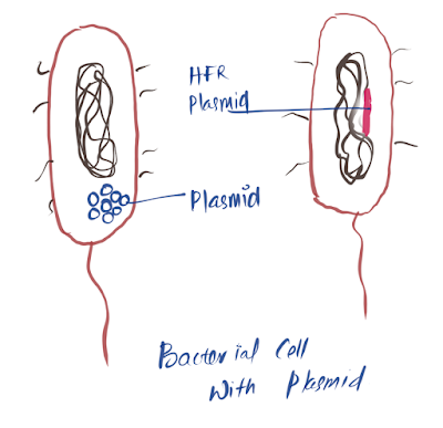
Plasmid:
-
These
are Extra chromosomal DNA.
-
Double
stranded, circular, self-replicating structures.
-
They
Gives unique character like
fertility, resistance etc to the cell.
-
Sometime
found attached to genome..
Called as Episomes or HFR plasmid
 |
| Plasmid-type |
Want to know about Shape size of Bacterial cell, Gram staining Click the link below :
Want to know about Ultra Structure of Bacterial cell Click the link below :
Inclusion Bodies.
•
These
are Nonliving structures of cytoplasm. Found in 2 conditions-
•
Free
- cyanophycean granules, volutin / phosphtate granules , glycogen granules
•
Cover
by very thin membrane – (2-4 nm nonolipid non unit protein) – gas vaculoles ,
carboxy some, sulphur garnules, PHB Granues (Poly 3 hydroxy butrate) storage of
carbon and energy..
•
On
the Basis of nature 3 types - gas vacuoles, inorganic inclusion and food
reserve.
Gas vacuoles
•
These
are Store of gas.
•
Mostly
in present in cyano bacteria and purple
bacteria.
•
Hexagonal,
hoollow, cylindrical gas vesicles can be seen.
•
Covered
by non lipid non unit protein membrane.
•
Which
Form many folds.
•
Help
in gaseous exchange, protect from radiation etc.
Inorganic inclusion
•
Examples
: Volutin, sulphur, iron, magnetite granules
•
These
are metachromatic granules-which show
different colour in basic dye.
•
Volutin-storage
of phosphate ( poly meta phosphate)
•
Sulphur
granules present in those bacteria which are living in sulphur rich medium.
•
Also
help in orientate themselves in geo magnetic field.
Food
reserve
•
In
BGA- cyanophycean starch or lipid globules or cyanophycin, or prtein granules
can be seen.
•
PHB Granules- Poly Beta hydroxy butrate granules present ( storage for Carbohydrate)
•
In
some Photo synthetic bacteria - Carboxy
somes present.
Flagella
•
Main
Locomotary organ.
•
1
to 7 micro meter in length, 0.02micro
meter in diameter (20nm)
•
Devided
to Basal body, hook and filament.
•
Basal
body- rod like swelling inside cell envelope- rings – 2 pairs
•
S & M ring in membrane and L & P ring in cell wall.
•
It
is just like a Motor system or rotator
system.
•
Hook
–is curved tubular structure.
•
Made
up off Flagellin protein.
•
9+2
arrangement seen in Flagella.
 |
| structure |
Pili or Fimbriae
•
These
are Non locomotary out growths.
•
Protein
present is – Pilin protein.
•
Pilli
– longer, few, thick , tubular ,
•
mostly
present in gram –ve bacteria (1-4 nos)
•
Help
in -attachment to
recipient cell during conjugation.
• Fimbriae- small bristle like, large number (300-400)
• help to attach in solid surface or to host or attachment with themselves.
 |
| Pili |
Want to know about Shape size of Bacterial cell, Gram staining Click the link below :
Want to know about Ultra Structure of Bacterial cell Click the link below :


Wonderful notes
ReplyDeleteThanks for appreciation.. stay connected👍👍
Delete